WP9 - Beyond HEP applications
WP Coordinators (ad interim):
• Dezso Varga
• Gabriele Croci
• Jona Bortfeldt
Contact: DRD1-WP9-contact@cern.ch
Participating institutes:
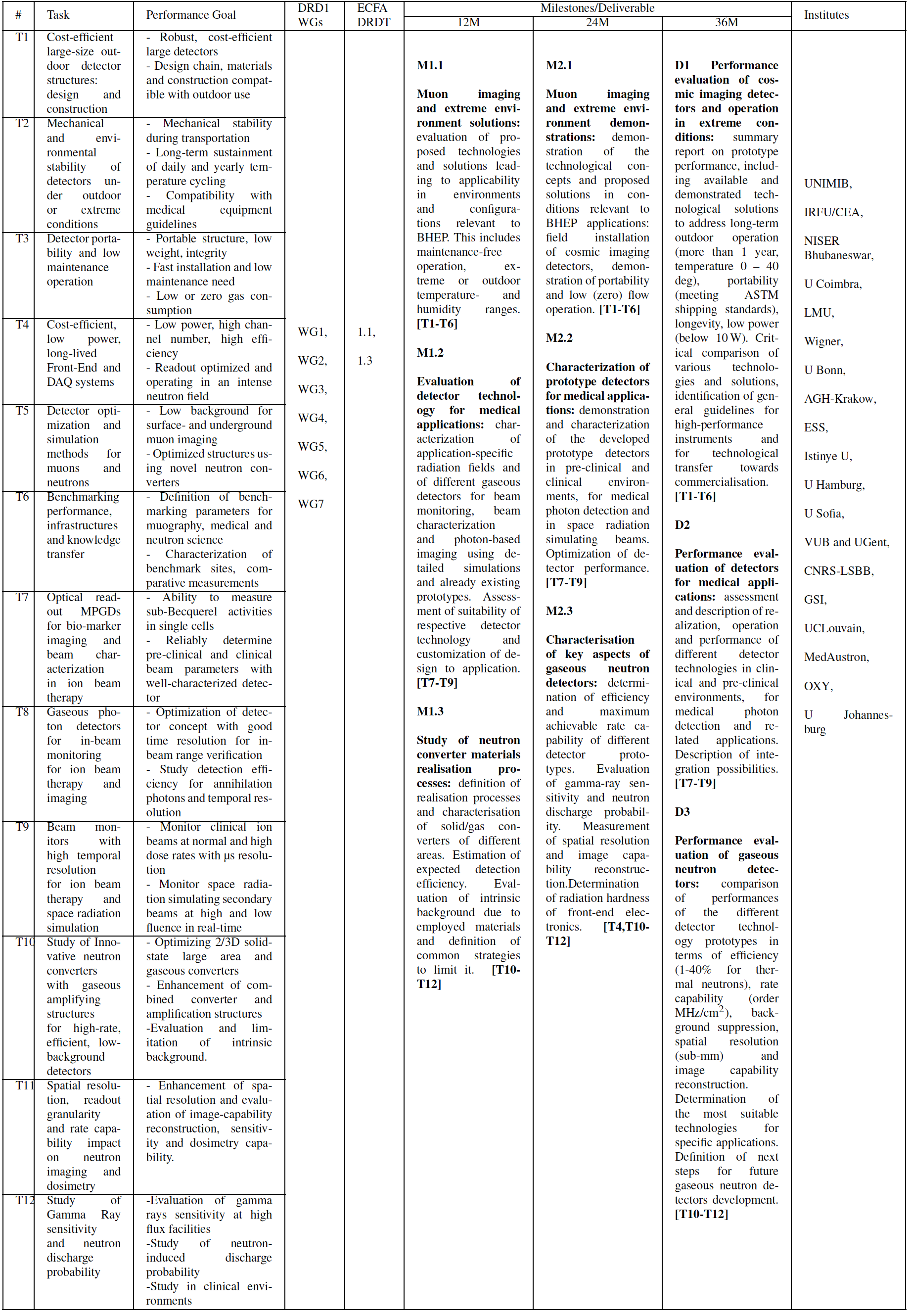
Università di Milano - Bicocca , University of Bonn, UCLouvain, LIP Coimbra, Istinye University, Wigner RCP, AGH University of Krakow, University of Hamburg, CEA/Saclay IRFU, LMU Munich, University of Sofia, MedAustron, VUB Brussel, LSBB, NISER, ESS, GSI, University of Johannesburg
DESCRIPTION OF THE WORK PACKAGE
Gaseous detector technologies are widely used in high energy and nuclear physics, which remain the driving force of cutting edge developments. The emergence of new technologies, as well as improvements leading to better performance, longer lifetime or radiation hardness and low-cost and mass production capabilities through collaboration with industries open the possibilities to apply such detectors outside HEP – many of those with high social and economic impact. The aim of the present Work Package is to fully exploit the achievements in other WP-s and to transfer knowledge and technologies to areas Beyond HEP. In fact such applications differ strongly from HEP in various aspects, including hostile or extreme conditions, very low level of maintenance, or strong requirements on operational stability, safety, gas emissions or structural stability. The present WP focuses on three larger sub-topics, namely cosmic muon imaging, medical applications and neutron science.
Muography
Cosmic muon imaging is a rapidly expanding interdisciplinary scientific field, its developments mainly driven by contemporary technological possibilities. The term “Muography” was coined recently, and includes classical applications such as imaging of cultural heritage – notably the “Great Void” discovery in the Khufu pyramid – or volcanoes, but finds use cases in mining industry, large buildings engineering, civil engineering and public safety, high resolution large object transmission tomography. The project includes a large fraction of the Muographers community, and aims at development of gaseous tracking detectors specifically for the challenging environments of field measurements. The project aims at practical, field-proven, high-TRL instrumentation. The new, fast and lightweight construction methods, heavy relying on industrial standard materials and techniques (e.g. 3D printing), need thorough field testing by experts of the relevant research fields, clearly outside the HEP community.
Medical Applications
Instrumentation development for medical physics applications, especially for photon and ion beam therapy and imaging, relies heavily on technology originally developed for high energy physics applications. An application in clinical systems however adds additional complexity to the instrumentation systems with respect to fail-safety, operational stability, operation in non-laboratory environments, portability and cost. Technology transfer into medical applications has in the past been very successful, however often carried by individual institutes. Dedicated instrumentation R&D in a potent collaboration will boost not only the technology transfer, but will enable considerable synergies in tailoring detection systems to pre-clinical and clinical
applications. Irradiation of moving targets, such as e.g. lung carcinoma or operation at high dose rates above 40 Gy/s in a total delivery time of around 100 ms, potentially reducing healthy tissue damage, require considerably faster beam monitoring systems in ion beam therapy. Typical motion mitigation strategies deliver the plan multiple times, resulting in up to ten times more spots to be delivered in the same timeframe. A conventional beam monitoring system takes a minimum amount of time per beam spot, typically some milliseconds, to safely monitor dose and position.To achieve sufficient delivery speed, the minimum times per spot have to be decreased to order of 100 µs. A functional detector for ultrafast beam-position monitoring will be desirable for research centres, clinics and companies working on both topics. With the background of future long-term space missions, in particular the already started ARTEMIS mission, radiation protection for astronauts, risk assessments and electronic hardness tests for on-board modules are becoming increasingly important. Intensity controlled raster point delivery of space radiation simulating secondary beams, produced in high energy synchrotrons, requires large-area and real-time beam monitors as a feed-back element in the beam delivery system. This is especially important as currently available systems become inaccurate for low fluences and larger fields.
Neutron Science
Neutron science experiments using fast or thermal neutrons are the core activity at spallation neutron sources and nuclear reactors and up to now detection systems have made great use of 3He-based gaseous detectors. During the last 10 years the 3He shortage and the massive use of this gas in applications linked to homeland security have determined an exponential rise of its price, preventing its use for research applications including present and future neutron sources (ILL, ESS, ISIS…). In the case of ESS, together with the need for replacing 3He, it is essential to develop high-rate neutron detectors made with low-background materials able to fully exploit the increased neutron flux of ESS relative to the present neutron sources . The situation called on one side for the development of large area, high-rate and neutron detectors with a comparable detection efficiency to those of 3He but with a price ideally not exceeding 500 k€/m2 and a spatial resolution between 1- 10 mm. On the other side, we want to develop low material budget beam monitors that can also provide real-time reconstruction of the 2D neutron profile. A series of MPGD based detectors have emerged in the last few years to cope with these requirements. Also applications linked to neutron detection in nuclear fusion experiments have driven the development Although MPGD-based detectors are mostly used to detect charged particles, they can be adapted (typically by using a customised cathode configuration), to detect fast and thermal neutrons In parallel also wire-based technology evolved in this direction (for example with the realisation of boron based straw tubes) as well as Resistive Plate Chamber technology. Several applications have already profited from these new developments but we think that in order to further progress a synergic action of all the research groups involved must be taken.
Drivers from applications
- Public safety and border control
- Mining industry
- Non-destructive imaging of sensitive objects
- Clinical photon and ion radiation therapy
- In-beam range monitoring in ion therapy
- Pre-clinical and clinical beam instriúmentation, also at ultra-high dose rates
- Low-fluence capable monitors for space radiation simulating secondary beams
- High Efficiency and high rate thermal neutron detectors
- Imaging with high rate fast and thermal neutrons
- Low-budget fast and thermal neutron beam monitors, beam profilers and beam loss monitors for safety applications
- Low background thermal epithermal and fast neutrons detectors
- Fast Neutron Spectroscopy
Common challenges:
- Portable and recirculating gas systems
- Sealed detectors or ultra-low gas consumption
- Operational stability in outdoor natural or extreme environments
- Cost efficient solutions for robust large detectors
- Very low maintenance level
- Neutron converters
- Efficiency
- Front-End electronics radiation hardness
- Low background materials
- Environmental-friendly gas mixtures
- Large Area granularity
- Sensitivity
- Physics applications (e.g neutron differential cross section studies)
- Low material budget
TASKS AND DELIVERABLES
T1: Cost efficient large size outdoor detector structures: design and construction
Muon imaging quality is directly related to detector size due to the low natural muon flux. Cost efficiency is highly relevant for applications, using robust detectors, therefore both aspects need to be considered simultaneously.
- D1: Demonstration of outdoor operability of large size detectors ( > 50cm) with various technologies, documentation of methods and design features leading to reduced cost without compromising performance.
T2: Mechanical and environmental stability of detectors under outdoor or extreme conditions
Environmental conditions for most muon imaging applications are drastically different from standard laboratory conditions experienced in HEP. Detector systems must be fully compatible with these outdoor parameters, and must reliably tolerate mechanical effects during transportation and installation. Beam instrumentation in clinical and related application is the integral element in beam delivery. Any failure or mis-measurement can have fatal consequences. Essential is thus the operational stability, independent of variable environmental conditions.
- D2: Performance demonstration of design options, assessment of conditions to qualify detectors for various applications (from arctic to hot climates, high humidity, strong pressure changes, high operational stability etc) and conditions during transportation (vibration, pressure change during air freight).
T3: Detector portability and low maintenance operation
Detector installation and operation time and effort must be kept at minimum for cost- and labour-efficient muon imaging measurements. This requires portability of the systems (modular structure, low weight, fast installation, reliable re-commissioning) as well as fast and reliable verification of proper functioning. Very low gas consumption is a specific requirement, or even running without gas supply in “sealed mode”. Similar requirements are equally important for the application of systems in clinical environments; Especially as typically no services such as cooling water and gas supply are available.
- D3: Demonstration of full portability, scalability by modular structure, as well as operation at low or zero gas flow during the data taking. Documentation of solutions to reduce installation, maintenance, and functional verification.
T4: Cost efficient, low power, long-lived Front-End and DAQ systems
Electronics components usually represent a large fraction of the cost, and consume most of the total supply power. Muography has characteristically low particle flux, which allows optimised designs to reduce both cost and power consumption while maintaining full efficiency within design resolution parameters. In case of neutron detector applications in intense neutron fields the study of front-end robustness to neutron radiation is paramount. All the
front-end electronics to be used with such detectors will have to be tested in specific facilities to determine the rate of SEE (single event effects).
- D4: Demonstration of functionalities of low power, low cost front-end and readout systems for high resolution gaseous muon imaging,cosmic ray instruments and neutron detectors.
T5: Detector configuration optimization and simulation methods for surface- and underground environments for muons and neutron experiments
Detector geometries must be adapted to the specific measurement conditions, which can include surface-based detectors with high all-sky background, or underground detectors installed into tight spaces (including boreholes). Dedicated simulations are mandatory during the measurement preparatory phase as well as evaluation of the data, addressing challenges specific to muon imaging, such as backgrounds and composition cosmic radiation. Development of modelling tools and techniques to optimise detector performance including the simulation of charge transport, signal induction and the influence of converting elements for neutron detection.
- D5: Documentation of optimization and simulation algorithms, demonstration of improved simulation methods and improved geometries leading to high resolution and background suppression.
T6: Benchmarking performance, infrastructures and knowledge transfer
Performance evaluation of emerging technologies, particularly comparing options, is notoriously difficult. Some infrastructures (such as LSBB or ESS) may be defined as benchmarking locations. Instrument development relies heavily on state-of-the-art HEP systems, a horizontal knowledge transfer is necessary from other WP-s, as well as other DRDs (particularly scintillators). Usage of common data format can strongly facilitate comparison of measurements. The task includes technology transfer preparation towards industrial partners as well as definition of reference instrumentation in clinical applications.
- D6: Definition of benchmarking performance parameters, instruments and infrastructures, documentation of benchmark performance parameters (efficiency, resolution, background rate, noise level, integrated acceptance). Characterization of various benchmark sites.
T7: Optical readout MPGDs for biomarker imaging and beam characterization in ion beam therapy
Optical readout MPGDs exploit the production of light in electron avalanches in the narrow amplification gap to register energy deposition in the active area of the device. They can be interfaced by cameras with high spatial resolution and by fast single-photon sensitive light detectors. As the gas gain and thus the amount of produced light is easily adjustable, the same device can be sensitive to single particles and photons or to intense beams. We propose to simulate, design and develop prototype detectors, suitable for individual gamma and charged particle detection as well as for ion beam characterization.
- D7.1: Demonstration of the capability to measure sub-Becquerel activities (3H and 14C) in single cells with an optical readout Micromegas prototype.
- D7.2: Development and characterization of an optical readout Micromegas device in pre-clinical and clinical particle beams.
T8: Gaseous photon detectors for in-beam monitoring for ion beam therapy and imaging
Ion beam therapy is not yet exploiting its full potential with respect to tumour targeting accuracy due to range uncertainties. They could be mitigated by determining during or shortly after each fractionated treatment session the accurate beam stopping position inside the patient. One possibility is to locate the beam-induced tissue activation by positron emission tomography. 511keV-sensitive RPCs, installed close to the patient couch, can enable accurate and fast delivery monitoring. Additional to range monitoring, gaseous photon detectors can be studied for use in other photon-based imaging modalities, such as imaging Positron Emission Tomography, SPECT, or for dosimetry applications.
- D8: Assembly of a prototype for annihilation photon detection and study of its parameters on a dedicated experimental setup after MC simulation to determine optimum design parameters.
T9: Beam monitors with high temporal resolution for ion beam therapy and space radiation simulation
Position and intensity of clinical ion beams are monitored at the beamline exit with thin O(0.5m²) beam monitor chambers. Gaseous detectors (ionisation chambers, MWPCs) are usually used for this purpose, due to their low ageing, stability and rate capability. For increasing treatment performance, monitoring and controlling future more intense clinical beams, especially in ultra-high dose rate conditions or in breathing cycle-timed delivery, or for real-time beam monitoring for space radiation simulation using raster scanning beam delivery however, the temporal resolution of current chambers is by two orders of magnitude too coarse. We propose to develop low-material budget beam monitor chambers, incorporating an MPGD amplification structure, rendering the device sensitive only to the rapidly moving ionisation electrons and to raster points with low number of particles. Adapting the currently used particle integrating readout electronics to switchable single particle sensitivity at 1MHz rate can enable individual particle imaging with the beam monitor. Report on the simulation, design and characterization of a prototype system with high temporal resolution for clinical beam monitoring with MHz temporal resolution and large dynamic range will be produced.
- D9.1: Report on the simulation, design and characterization of a prototype system with high temporal resolution for clinical beam monitoring with MHz temporal resolution and large dynamic range.
- D9.2: Report on the design, characterization and optimization of a full-scale, low-material budget prototype for space radiation-like beam monitoring. Configurable for particles from carbon to uranium in a range from 100 MeV/u to 2 GeV/u, with spatial resolution better 0.5mm and fast feedback <10µs to the beam control system.
T10: Innovative neutron converter geometries in combination with gaseous amplifying structures for high rate, efficient, low background detectors
Study and optimization of the realisation processes of (2D or 3D) solid-state neutron converters (eg. based on B, Li, Gd for thermal and slow neutrons or U-238 and other fissile materials and hydrogenated for fast neutrons) towards the production of uniform, reliable, stable and large area (around m2) neutron converters. Beyond solid state we propose also to study gaseous neutron converters (eg. N). Enhancement of solid-state converter geometries (especially 3D) to improve detector efficiency. Study of the sensitivity taking into account the intrinsic resolution. Detector studies and optimization (e.g. amplification and charge evacuation) to evaluate the compatibility of present and future neutron converter generation with amplifying structures. Enhancement of combined neutron converter and amplification structures realisation. Evaluation of intrinsic background due to employed materials in the detectors and implementation of common strategies to limit it.
- D10.1: Report on realisation processes and characterization of 2D or 3D solid converters of different areas to determine chemical and physical properties.
- D10.2: Report on efficiency measurements of different detectors realised by coupling solid state or gaseous converters to various amplification structures
- D10.3: Report on evaluation of intrinsic background due to employed materials in the detectors and on common strategies strategies to limit it
T11: Spatial resolution, readout granularity and rate capability impact on neutron imaging and dosimetry
Enhance spatial resolution of neutron detectors by optimising readout granularity and develop readout solutions which can improve spatial resolution. Evaluation of image-capability reconstruction for imaging applications, sensitivity and dosimetry in clinical environments.
- D11: Report on spatial resolution and image capability reconstruction with different detectors
T12: Study of Gamma Ray sensitivity and neutron discharge probability
Evaluation of gamma rays sensitivity of gaseous neutron detectors (gamma ray field associated with neutrons has an average energy of 1 MeV) by using high flux facilities like nTOF and GIF++ at CERN or other accelerator facilities (e.g clinical accelerators). Detailed study of neutron induced discharge probability will be performed. These studies are relevant also for beam monitors for clinical environments, especially at high dose rates or in high-gain single particle sensitive operation.
- D12: Report on evaluation of gamma ray sensitivity and neutron discharge probability of different detectors subjected to high dose rate of gammas and neutrons.